
Figure 6.
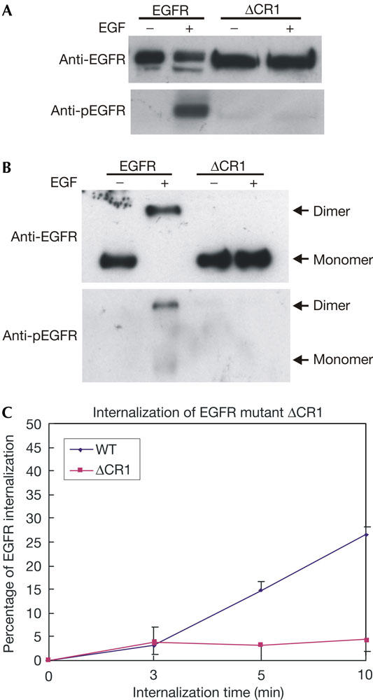
Inhibition of epidermal growth factor-induced internalization of ΔCR1 in BaF/3 cells stably expressing ΔCR1. BaF/3 cells stably expressing epidermal growth factor receptor (EGFR) or ΔCR1 were stimulated with EGF (100 ng/ml) for 15 min. (A) The cells were lysed and the protein samples were immunoblotted with anti-EGFR and anti-phospho-EGFR (pEGFR) antibodies. (B) The cells were crosslinked with bis-(sulphosuccinimidyl suberate) (BS3) and lysed. The protein samples were immunoblotted with anti-EGFR and anti-pEGFR antibodies. (C) Quantitative analysis of EGFR internalization in BaF/3 cells by flow cytometry. BaF/3 cells stably expressing EGFR or ΔCR1 were stimulated with EGF (100 ng/ml) for the indicated time and the internalization of ΔCR1 was analysed by flow cytometry. WT, wild type.