Figure 1.

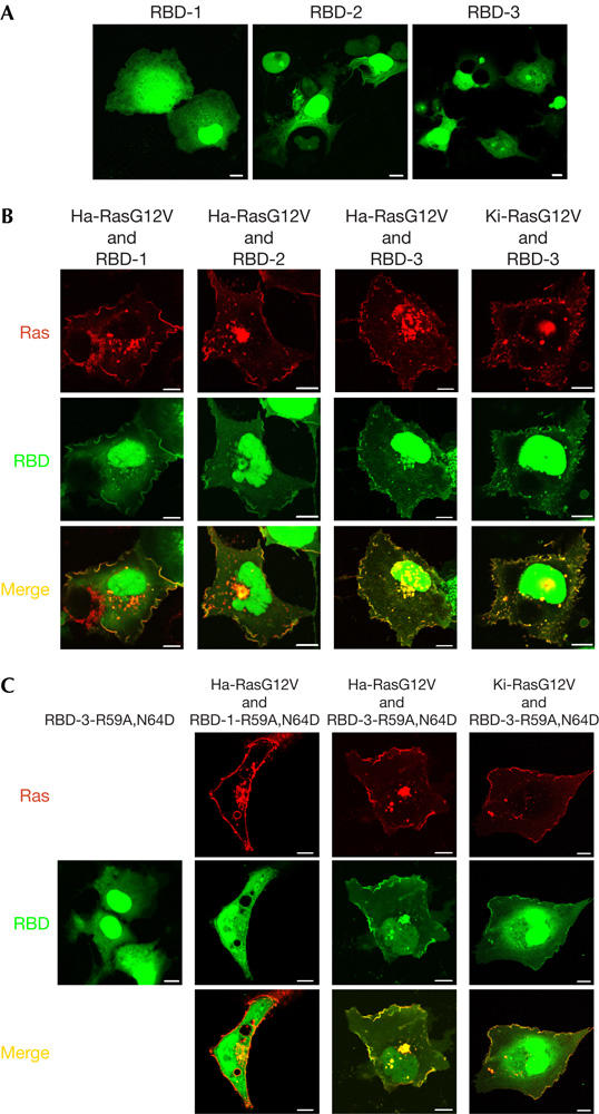
Cellular effects and distribution of oligomerized RBD polypeptides. (A) Distribution of GFP–RBD-1/2/3 in serum-starved COS-7 cells. More than 90% of transfected cells showed the described phenotype. GFP, green fluorescent protein; RBD, Ras-binding domain of the Ras effector c-Raf. (B) COS-7 cells were transfected with DsRed-tagged Ha-RasG12V or Ki-RasG12V in combination with GFP–RBD-1/2/3, as indicated. Cells were serum starved and imaged alive. Yellow pseudo-colour marks colocalization. (C) GFP–RBD-1-R59A,N64D or GFP–RBD-3-R59A,N64D was co-transfected with DsRed-Ha-RasG12V or DsRed-Ki-RasG12V, as indicated. Protein distribution was imaged confocally. Scale bars, 10 μm.
