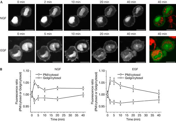
Figure 4.

Ras activation in PC12 cells proceeds at the plasma membrane but not at the Golgi. (A) Serum-starved PC12 cells transfected with GFP–RBD-3 were stained with the Golgi marker BODIPY-TR-C5-ceramide, challenged with 50 ng/ml nerve growth factor (NGF) or epidermal growth factor (EGF) and imaged alive. White and green, GFP–RBD-3; red, BODIPY-TR-C5-ceramide. GFP, green fluorescent protein; RBD, Ras-binding domain of the Ras effector c-Raf. Scale bars, 10 μm. (B) Quantification of fluorescence associated with the plasma membrane (PM) and Golgi for the experiments shown in (A), plotted as the ratio of mean pixel fluorescence intensity at the PM or Golgi to cytosol versus time (mean±s.e.m.; NGF: n=6; EGF: n=9). The value for the unstimulated cell was set as 1. For a description of the quantification procedure, see supplementary Fig 8 online.
