Figure 5.

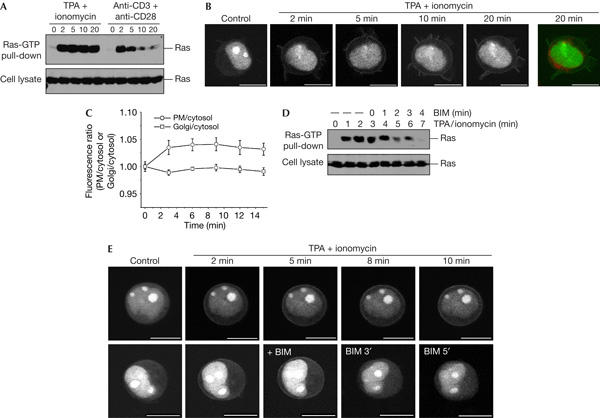
12-O-tetradecanoylphorbol-13-acetate/ionomycin activation of Ras in Jurkat cells does not occur at the Golgi. (A) Serum-starved Jurkat cells were challenged with 150 nM 12-O-tetradecanoylphorbol-13-acetate (TPA) plus 500 ng/ml ionomycin or anti-CD3 plus anti-CD28 (5 μg/ml each), to activate the T-cell receptor, lysed and processed in a Ras-GTP pull-down assay. (B) Serum-starved Jurkat cells expressing GFP–RBD-3-R59A were stained with BODIPY-TR-C5-ceramide, challenged with TPA/ionomycin and imaged alive. White and green, GFP–RBD-3-R59A; red, BODIPY-TR-C5-ceramide. GFP, green fluorescent protein; RBD, Ras-binding domain of the Ras effector c-Raf. (C) Quantification of fluorescence associated with the plasma membrane and Golgi for the experiment shown in (B) (n=8). Refer to legend to Fig 4B. (D) Serum-starved Jurkat cells were stimulated with TPA/ionomycin as before. At 3 min after stimulation, the cell suspension was made up to 5 μM bisindolylmaleimide I (BIM). After the indicated time points, samples were processed in a Ras-GTP pull-down assay. (E) Serum-starved Jurkat cells expressing GFP–RBD-3-R59A were challenged with TPA/ionomycin and imaged confocally. In the lower panel, the medium was made up to 5 μM BIM at the indicated time point. Scale bars, 10 μm.
