
Figure 1.
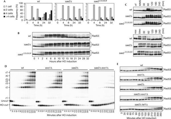
Response to a single irreparable double-strand break in sae2Δ cells. (A) YEP+raf G1-arrested cell cultures of wild-type (wt) JKM139 and isogenic sae2Δ and sae22,5,6,8,9 strains were spotted on galactose-containing plates incubated at 30°C (time zero). At the indicated time points, 200 cells for each strain were analysed to determine the frequency of single cells and of cells forming microcolonies of two, four or more than four cells. (B) Galactose was added at time zero to wild-type JKM139 and isogenic sae2Δ and sae22,5,6,8,9 cell cultures exponentially growing in YEP+raf. Protein extracts from aliquots withdrawn at the indicated times were analysed by western blot with anti-Rad53 antibodies. (C) Wild-type W303 and isogenic sae2Δ and sae22,5,6,8,9 cell cultures arrested in G1 with α-factor (αf) or in G2 with nocodazole (noc) were incubated for 15 min with methylmethane sulphonate (MMS, 0.02%) or bleomycin (bleo, 10 mU/ml), respectively, and then released in YEPD. Protein extracts from samples taken at the indicated times were analysed by western blot with anti-Rad53 antibodies. (D) YEP+raf nocodazole-arrested cell cultures of wild-type JKM139 and isogenic exo1Δ, sae2Δ, sae2Δ exo1Δ and sae2Δ tel1Δ strains were transferred to YEP+raf+gal in the presence of nocodazole at time zero. Genomic DNA prepared from aliquots taken at the indicated times was digested with SspI and separated on alkaline agarose gel. Gel blots were hybridized with a single-stranded RNA probe specific for the MAT locus, which shows HO-cut and uncut fragments of 0.9 and 1.1 kb, respectively. As depicted in supplementary Fig S2 online, 5′-to-3′ resection progressively eliminates SspI sites located 1.7, 3.5, 4.7, 5.9, 6.5, 8.9 and 15.8 kb centromere-distal from the HO-cut site, producing larger SspI fragments (r1–r7) detected by the probe. The kinetics of resection product accumulation in sae2Δ tel1Δ cell cultures (not shown) was undistinguishable from that of sae2Δ cells. (E) Protein extracts from samples taken at the indicated times during the experiment in (D) were analysed by western blot with anti-Rad53 antibodies.