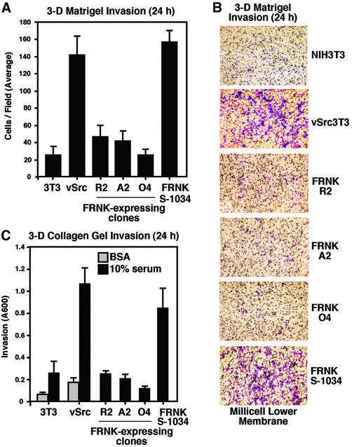
Fig. 2. FRNK inhibits v-Src-stimulated three-dimensional cell invasion. (A) Matrigel (30 µg) invasion assays were performed with the indicated cells for 24 h using a serum stimulus in the lower chamber. Values are means ± SD from four experiments. No cell invasion was detected in the presence of BSA. (B) Representative images (60×) of the lower porous membrane surface from Matrigel invasion assays. Crystal violet-stained cells can be distinguished from the 8 µm membrane pores. (C) Cell invasion through polymerized collagen type I was assayed with the indicated cells for 24 h using a serum stimulus in the lower chamber. Values are means ± SD from two independent experiments. Random invasion activity was assayed in the presence of BSA.

An official website of the United States government
Here's how you know
Official websites use .gov
A
.gov website belongs to an official
government organization in the United States.
Secure .gov websites use HTTPS
A lock (
) or https:// means you've safely
connected to the .gov website. Share sensitive
information only on official, secure websites.
