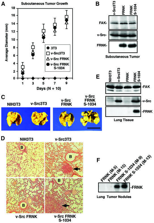
Fig. 5. FRNK inhibits experimental metastasis but not v-Src-stimulated solid tumor growth. (A) Athymic nude mice were injected subcutaneously with the indicated cells and, after 10 days, visible tumor size was measured with calipers. Values are means ± SD from two experiments with eight animals per group. NIH-3T3 (diamonds), v-Src3T3 (open squares), v-Src FRNK (open triangles) and v-Src FRNK S-1034 (filled circles). (B) Protein lysates from the indicated subcutaneously grown tumor tissue were analyzed by FAK (top), v-Src (middle) or HA tag blotting (lower). (C) Representative Bouin’s-fixed lungs from mice 4 weeks after injection with the indicated cells. Multiple tumor nodules on the lung surface can be seen in v-Src3T3- and v-Src FRNK S-1034-injected animals. The scale bar is 1 cm. (D) Representative H&E-stained mouse lung sections from animals injected with the indicated cells. Metastatic tumor cells (arrows) and blood vessels (B) are indicated. (E) Blotting analyses of lung tissue after 4 weeks from animals injected with the indicated cells. FAK blotting was performed on protein lysates (top), v-Src was detected by immunoprecipitation and blotting (middle), and FRNK was detected by HA tag immunoprecipitation and blotting (lower). (F) HA tag blotting analysis was performed on protein lysates of lung tumor nodules from either v-Src FRNK- or v-Src FRNK S-1034-injected mice. The M number is used to identify mice.

An official website of the United States government
Here's how you know
Official websites use .gov
A
.gov website belongs to an official
government organization in the United States.
Secure .gov websites use HTTPS
A lock (
) or https:// means you've safely
connected to the .gov website. Share sensitive
information only on official, secure websites.
