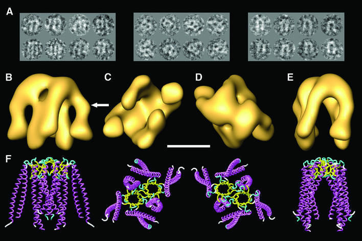
Fig. 1. Three-dimensional reconstruction of human PFD. (A) A gallery of negatively stained images similar to those used for the three-dimensional reconstruction of PFD. The three groups of images shown are archetypes of the three main views, which are orthogonal. (B–E) Several views of the three-dimensional reconstruction of human PFD. (F) Similar views of the atomic structure of the archaeal homolog of PFD (MtGimC from M.thermoautotrophicum; Siegert et al., 2000); PDB accession code 1FXK. The arrow points to a kink in the eukaryotic structure of PFD that is found reproducibly in all the three-dimensional reconstructions generated, and which is not present in the atomic structure of the archaeal PFD. Scale bar = 50 Å.

An official website of the United States government
Here's how you know
Official websites use .gov
A
.gov website belongs to an official
government organization in the United States.
Secure .gov websites use HTTPS
A lock (
) or https:// means you've safely
connected to the .gov website. Share sensitive
information only on official, secure websites.
