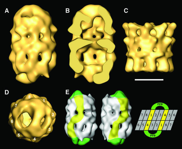
Fig. 4. Three-dimensional reconstruction of the CCT:PFD symmetric complex. (A) Side view of the CCT:PFD symmetric complex. (B) A section along the longitudinal axis of the same volume. (C) Side view of the three-dimensional reconstruction of apo-CCT (Llorca et al, 2000). (D) Top view of the three-dimensional reconstruction showing the 1,4 interaction between PFD and the CCT subunits. (E) A scheme showing the interaction of the two PFD oligomers with the CCT subunits of the two rings. The two volumes on the left represent two opposing views of the CCT:PFD complex. The PFD oligomers are highlighted in green and the CCT subunits interacting with PFD are shown in yellow. The topology of the CCT subunits depicted on the right is for illustrative purposes only, and the numbers have the sole purpose of discriminating among the subunits. Scale bar = 100 Å in (A–D).

An official website of the United States government
Here's how you know
Official websites use .gov
A
.gov website belongs to an official
government organization in the United States.
Secure .gov websites use HTTPS
A lock (
) or https:// means you've safely
connected to the .gov website. Share sensitive
information only on official, secure websites.
