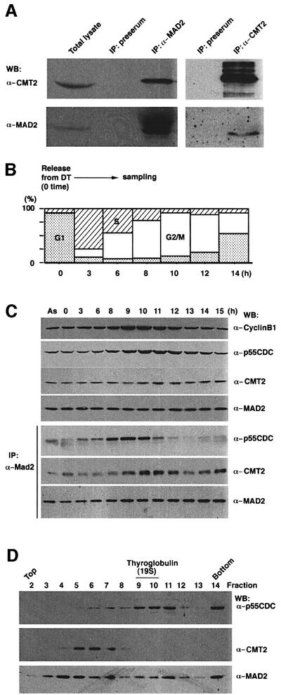
Fig. 3. CMT2–MAD2 and p55CDC–MAD2 complexes. (A) Immuno precipitation was performed with the antibody to MAD2 (lane 3) or antibody to CMT2 (lane 5) with extracts prepared from an asynchronous HeLa cells culture. The precipitates were run on SDS–PAGE and analyzed by western blotting. Total lysates used for immunoprecipitation (lane 1) and precipitates by pre-immune serum (lanes 2 and 4) were also analyzed. (B) Cell culture, blocked by double thymidine (DT), was released at time 0 and sampled for (C). Synchronization of the culture was confirmed by FACS analysis. (C) Cell cycle analysis. Cell extracts were prepared at indicated time points after the release and processed for western blotting (upper three panels), or immunoprecipitation with the antibody to MAD2 followed by western blotting (lower three panels). Extracts prepared from an asynchronous culture (AS) were also examined in the same way. (D) Glycerol gradient centrifugation. Mitotic HeLa cell extracts were subjected to glycerol gradient centrifugation. Each fraction was run on SDS–PAGE and processed for western blotting.

An official website of the United States government
Here's how you know
Official websites use .gov
A
.gov website belongs to an official
government organization in the United States.
Secure .gov websites use HTTPS
A lock (
) or https:// means you've safely
connected to the .gov website. Share sensitive
information only on official, secure websites.
