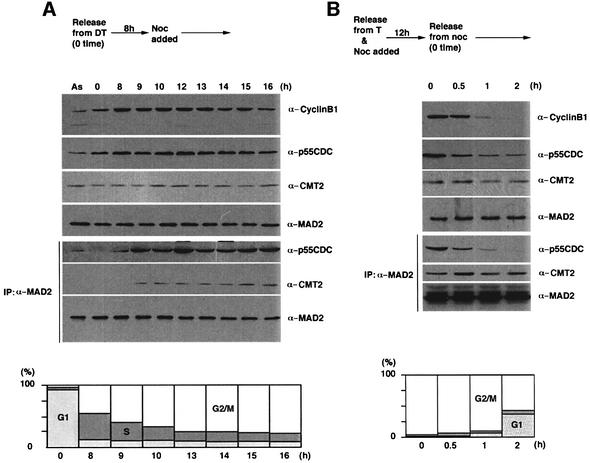
Fig. 4. Transition from p55CDC–MAD2 to CMT2–MAD2. (A) HeLa cells were arrested by double thymidine (DT) block and released at time 0. Nocodazole was added at 8 h after the release. Extracts were prepared at indicated time points and processed for western blotting (upper three panels), or immunoprecipitation with the antibody to MAD2 followed by western blotting (lower three panels). Extracts prepared from an asynchronous culture (AS) were also examined in the same way. FACS analysis was performed to monitor cell cycle progression. (B) HeLa cells were arrested by a single thymidine (T) block and released into the media containing nocodazole. After a 12 h incubation, the cells were released into the drug-free media (time 0). Cell extracts were prepared at indicated time points for western blotting (upper three panels), or immunoprecipitation with the antibody to MAD2 followed by western blotting (lower three panels). FACS analysis was performed to monitor cell cycle progression.

An official website of the United States government
Here's how you know
Official websites use .gov
A
.gov website belongs to an official
government organization in the United States.
Secure .gov websites use HTTPS
A lock (
) or https:// means you've safely
connected to the .gov website. Share sensitive
information only on official, secure websites.
