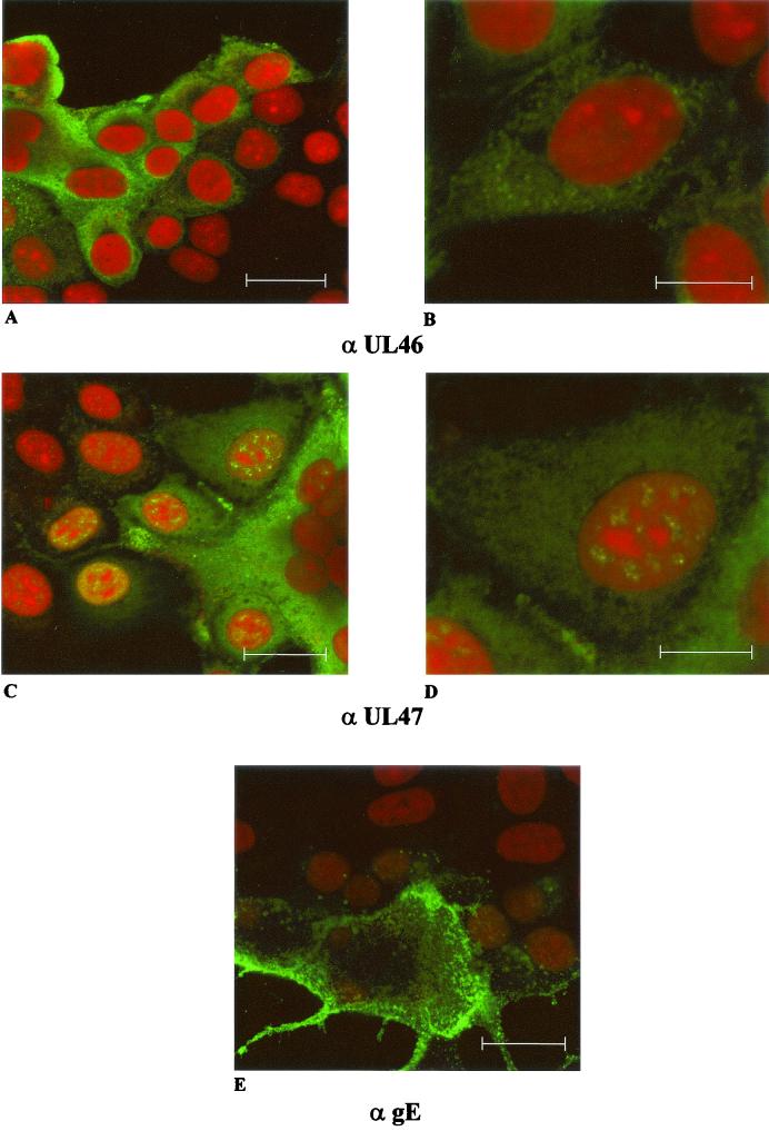
FIG. 4.
Intracellular localization of PrV UL46 and UL47 proteins. Immunofluorescence analysis by confocal laser scan microscopy of RK13 cells infected with PrV-Ka under plaque assay conditions was performed by using monospecific antisera against the UL46 (upper row) or UL47 proteins (middle row) or a monoclonal antibody against gE (bottom). For UL46 and UL47 detection, two different magnifications are shown (bars represent 25 μm in left panels and 50 μm in right panels). Red, propidium iodide stain; green, reactivity of specific antibodies.

