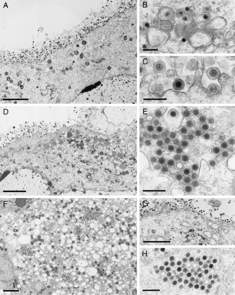
FIG. 6.
Electron microscopy of mutant virus-infected cells. RK13 cells were infected with PrV-ΔUL46 (A to C), PrV-ΔUL47 (D and E), or PrV-ΔUL46/47 (F to H) and analyzed by electron microscopy 16 h after infection. (A to C) Unimpaired virion morphogenesis in the absence of the UL46 protein including secondary envelopment in the cytoplasm (B) as well as numerous extracellular virions (A and C) are shown. (D) Overview of a PrV-ΔUL47-infected cell demonstrating that besides all stages of normal virion morphogenesis, intracytoplasmic aggregations of capsids can be observed (shown at higher magnification in panel E). Similar aggregations were also observed in PrV-ΔUL46/47-infected cells (F to H). Bars, 3 μm in panels A, D, F, and G and 300 nm in panels B, C, E, and H.

