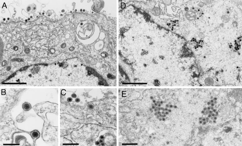
FIG. 7.
Electron microscopy of infected RK13-UL46/47 cells. Transcomplementing RK13-UL46/47 and normal RK13 cells were infected with PrV-ΔUL47 and analyzed by electron microscopy 16 h after infection. Panels A to C show unimpaired virion formation on complementing cells, whereas panels D and E demonstrate, in a parallel assay, the formation of capsid aggregates in the cytoplasm of noncomplementing cells. Bars, 1.5 μm in panels A and D, 100 nm in panel B, and 500 nm in panels C and E.

