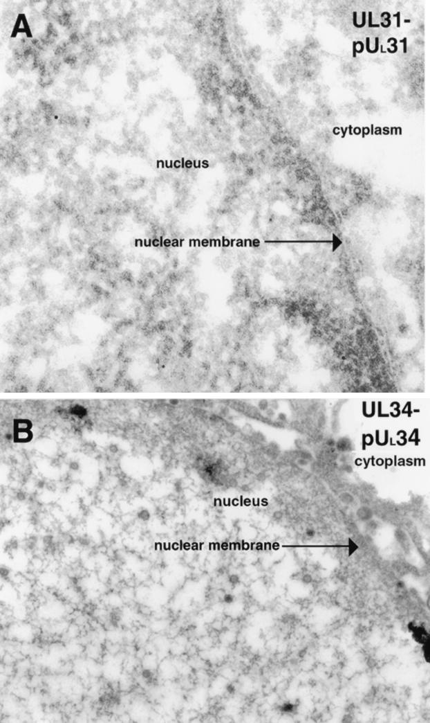
FIG. 2.
Scanned digital electron micrograph of UL31-null mutant virus-infected thin sections of Vero cells (A) or UL34-null mutant virus-infected thin sections of Vero cells (B) harvested and fixed late in infection (14 to 18 hpi). In panel A, thin sections were probed with UL31-specific rabbit antisera. The cytoplasm, nuclear membrane, and nucleus are all labeled. Original magnification of panel A, ×45,000. In panel B, thin sections were probed with UL34 protein-specific IgY antibodies. Original magnification of panel B, ×45,000.

