FIG. 1.
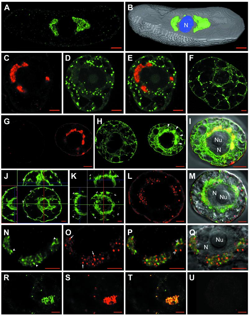
Confocal immunomicroscopy on healthy or GFLV-infected T-BY2 protoplasts. The cell lines used were: wild type (A and B, N to U), transgenic cells expressing GFP targeted to the Golgi (C to F) or to the ER (G to M). Mock-inoculated and infected protoplasts were harvestedat 24 hpi (R to U) or 48 hpi (A to Q). To monitor the synthesis of viral RNA, cells were fed at 24 hpi with BrUTP in the presence of actinomycin D and incubated for 6 additional hours to pulse-label the viral RNA (R to U). Cells were fixed with glutaraldehyde, permeabilized, quenched with NaBH4, and treated with primary antibodies, followed by treatment with secondary antibodies coupled to fluorochromes (Ab1 and Ab2). Immunolabelings were performed with anti-VPg/A488 (A, B, N, P, and Q), anti-VPg/A568 (C, E, G, and I), anti-BrdU/A488 (R and T), and anti-dsRNA/A568 (L, M, O to Q, and S to U). Panels G and H show fields containing two adjacent cells; the left cell escaped infection and represents a healthy cell, whereas the right cell is infected and shows condensed ER (H; large full arrowheads). Panels J and K are orthogonal projections of two series of optical sections through a healthy cell with a typical cortical ER (J; single arrowheads) and an infected cell showing a depletion of the cortical ER (K; single arrowheads). Merged pictures were obtained by combining the green and red channels (E = C + D; I = G + H, right cells; M = K + L; P and Q = N + O; and T = R + S). The VPg-containing perinuclear aggregates and punctate dsRNA labeling are indicated in panel N by full arrowheads and in panel O by arrows. Colocalization between VPg and dsRNA labeling are designated by double arrowheads in panel P. Superposition of differential interference contrast in panels I, M, and Q allowed visualization of the cell content and, particularly, the nucleus. N, nucleus; Nu, nucleolus. Panels F, J, and U correspond to healthy cells. Bars, 5 μm.

