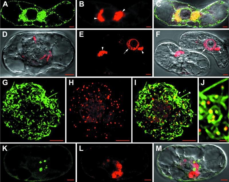
FIG. 6.
Confocal immunomicroscopy on healthy or GFLV-infected T-BY2 protoplasts. Cells were from transgenic lines expressing GFP in the ER (A, B, and C) or from wild type (D to M). Cells were harvested at 48 h p.i. and treated as in Fig. 1. Labelings were done with anti-CP/A568 (B, C, D, E, F, H, I, J, L, and M) or anti-MP/A488 (G, I, J, K, and M). Panels C, I, and M are merged pictures from panels A + B, G + H, and K + L, respectively. Differential interference contrast observations were superimposed in panels C, D, F, and M for visualization of the cell and its nucleus. Panel J is a 2.8-fold magnification of the area indicated in panel I. The arrowheads in panels B, C, E, and F indicate the perinuclear viral compartment. The arrows in panels E and F indicate the CP labeling in the nucleoplasm. The boxed region in panel I is shown enlarged in panel J. Bar, 5 μm except in panel J (1 μm).

