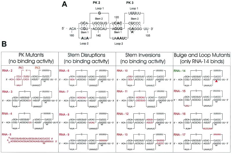
FIG. 7.
Fine analysis of eEF1A binding to PK2-PK3. (A) Primary and secondary structures of the wild-type sequence (RNA-1) of the PK2-PK3 region showing the stems and loops involved in PK formation. Conserved nucleotides are in bold. (B) Maps of the mutants analyzed. The modified sequences or structures are shown in red. Construct names (RNA-2 to RNA-17) are in red or green to indicate no binding activity or normal binding activity, respectively.

