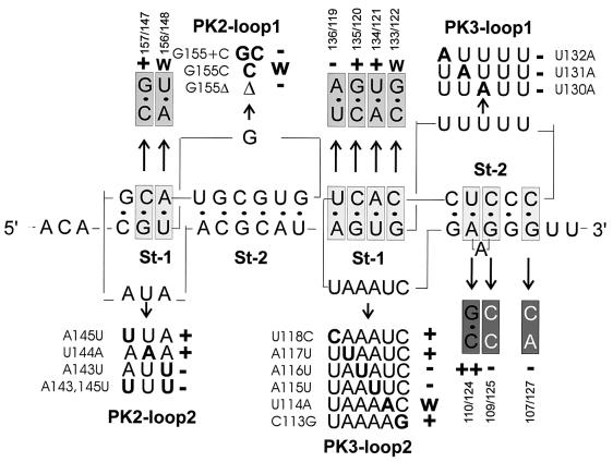
FIG. 8.
Analysis of single mutants in the loops and base pair mutants in the stems of PK2 and PK3 affecting eEF1A binding. Shown are primary and secondary structures of the region, with mutants analyzed indicated. Mutated nucleotides in the loops are shown in bold, and their effects in the UV cross-linking assay are indicated by + (wild-type-like), w (weakened binding), and − (loss of binding activity). Analyzed base pairs (boxed) were exchanged for flipped base pairs (light grey), strengthened base pairs (dark grey), or unpaired sequences (white on grey). Results are indicated as + (active), ++ (strongly active), w (weakly active), and − (inactive).

