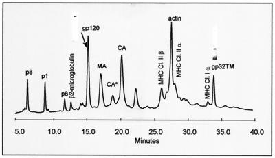
FIG. 1.
HPLC analysis of sucrose density gradient-purified SIVMne(E11S). After purification, virions were disrupted in 8 M guanidine-HCl and separated by HPLC. Peaks were detected at 280 nm. Proteins were eluted from the column with an acetonitrile gradient as described in Materials and Methods and identified by SDS-PAGE, immunoblot, mass spectrometry, protein sequencing, and amino acid analysis. Protein purity of SU(gp120) and TM(gp32) peaks is shown as insertions on the HPLC profile. Bands were visualized by Coomassie staining. Two UV-absorbing peaks (labeled CA* and CA) were found to contain highly purified monomeric Gag p28CA protein. Subsequent analysis showed that following reduction with 2-mercaptoethanol, the protein in peak CA* eluted as CA, indicating that CA* contained a form of p28CA with at least one internal disulfide bond.

