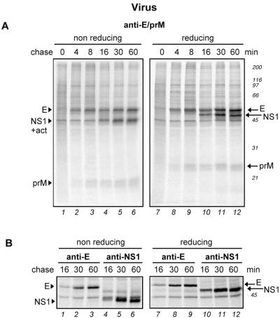
FIG. 2.
Folding of TBE virus envelope proteins in virus-infected mammalian cells. COS-1 cells were infected with TBE virus strain Neudoerfl at a multiplicity of infection of ca. 1. At 24 h postinfection, the cells were pulse-labeled with 35S-labeled methionine-cysteine for 2 min and chased for 0 to 60 min. This was followed by immunoprecipitation of the postnuclear supernatants with a polyclonal antiserum recognizing both prM and E (A) or MAbs specific for E and NS1, as indicated (B). The immunoprecipitates were analyzed by nonreducing and reducing SDS-PAGE (12% gel for panel A and 7.5% gel for panel B) and exposed for autoradiography. Positions of the individual proteins are marked at the side. In most of the gels, a nonspecific band was detected at ca. 45 kDa comigrating with NS1 under nonreducing conditions (+act) that probably corresponds to labeled actin. Molecular size standards are indicated on the right in kilodaltons.

