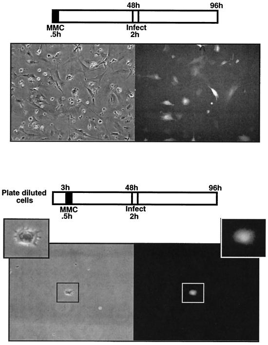
FIG. 5.
Infection of mitomycin C-arrested DF-1 cells with ASV. In the top panels, DF-1 cells were treated with mitomycin C for 0.5 h. After 48 h, cells were infected with ASVA-CMVEGFP and examined for GFP expression 48 h postinfection. Phase-contrast (left) and fluorescence (right) images (GFP) are shown. The bottom panels are the same as the top panels, except that DF-1 cells were monodispersed prior to mitomycin C treatment such that infection of individual cells could be observed. Shown is an arrested premitotic cell expressing GFP.

