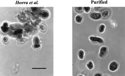
FIGURE 1.
Microscopy of isolated nuclei. Isolated HeLa cell nuclei, prepared either by the method of Iborra et al. (2001) or by the modified procedure, were mixed with an equal volume of 0.5% Azure C in 0.25 M sucrose (Busch 1967) and examined by phase-contrast microscopy. The scale bar is 10 μm.

