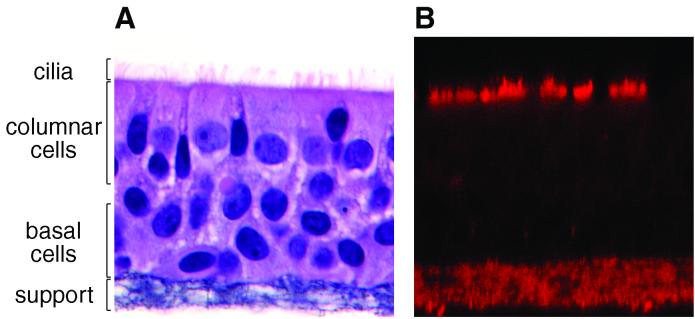
FIG. 1.
Cell morphology and KS expression at the apical ciliated surfaces of WD HAE cell cultures. (A) Light micrograph of a cross section of a WD HAE culture grown at an ALI on a semipermeable membrane support for 4 weeks. Under these conditions, pseudostratified mucociliary epithelial cell morphology was generated. The cells were counterstained with hematoxylin and eosin. (B) Confocal fluorescent optical section of a live WD HAE culture exposed to an antibody specific for KS and detected with a secondary antibody conjugated to Texas Red. Note that KS serves as a marker for ciliated columnar epithelial cells at the apical surface of the culture and that the permeable support, a 10-μm-deep layer underlying the basal epithelial cells, displays non-KS-specific autofluorescence. Original magnification, ×100.

