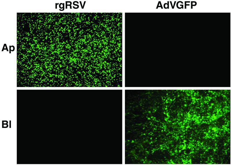
FIG. 2.
Comparison of the abilities of rgRSV and AdVGFP to infect the apical (Ap) versus the basolateral (Bl) surfaces of WD HAE cultures. rgRSV (7 × 10⁶ PFU; MOI, ∼20) or AdVGFP (108 PFU; MOI, ∼300) was applied to either the apical or basolateral surface of the cultures as detailed in Materials and Methods. Twenty-four hours later, the cultures were analyzed en face for GFP expression by fluorescence photomicroscopy. Original magnification, ×10.

