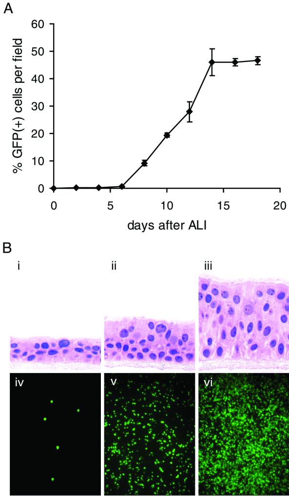
FIG. 5.
Susceptibility of HAE cultures to rgRSV infection as a function of the differentiation state of the culture. (A) Freshly plated cells were grown to confluence to represent a PD cell type and allowed to differentiate with time. On the indicated days following establishment of an ALI, replicate cultures were inoculated with rgRSV (7 × 10⁶ PFU), and the percentage of GFP-positive cells was quantitated by fluorescence photomicroscopy 24 h later. Each datum point represents the mean of three independent measurements ± standard error of the mean. (B) Representative photomicrographs of the differentiation status of HAE cultures on day 2 (i), day 8 (ii), and day 14 (iii) after initiation of an ALI. Note the abundant ciliated cells on day 14. The cells were counterstained with hematoxylin and eosin. Also shown are en face fluorescence photomicrographs of corresponding cultures expressing GFP 24 h after inoculation with rgRSV on day 2 (iv), day 8 (v), and day 14 (vi). Original magnifications, ×100 (light) and ×10 (fluorescence).

