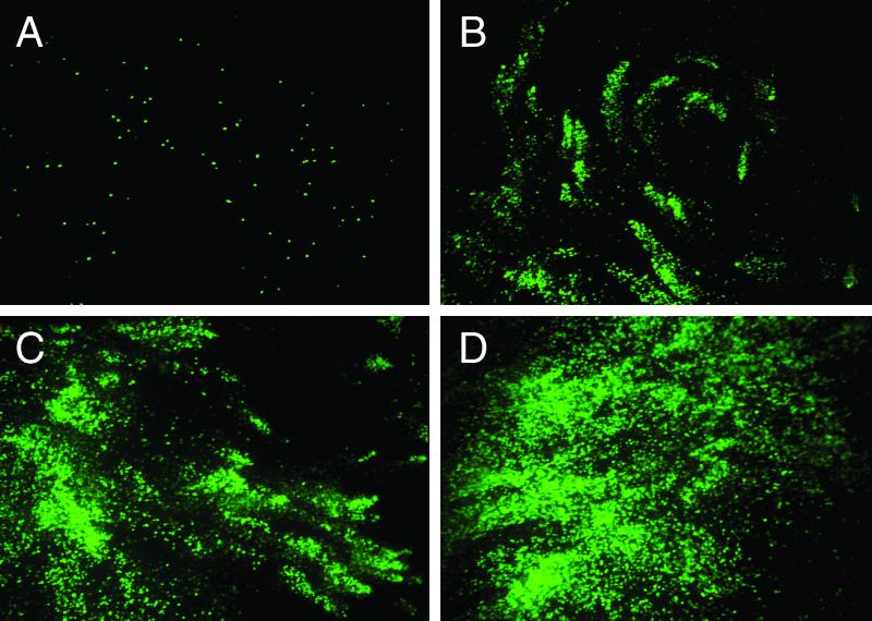
FIG. 7.
Spread of rgRSV infection with time in WD HAE cultures. The apical surfaces of cultures were inoculated with a low titer of rgRSV (7 ×10³ PFU) to achieve a submaximal number of cells expressing GFP at 24 h. Infection was then allowed to proceed over 4 days, and GFP expression was examined en face by fluorescence photomicroscopy on days 1 (A), 2 (B), 3 (C), and 4 (D) postinoculation. Note the counterclockwise circular spread of rgRSV infection by day 2 (B) and the increased number of rgRSV-infected cells by day 4. Original magnification, ×10.

