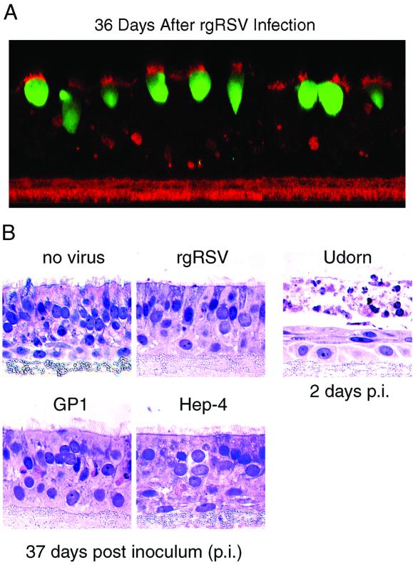
FIG. 9.
Lack of RSV-specific obvious cytopathology in WD HAE cells. (A) Confocal optical section of rgRSV-mediated GFP expression 36 days after apical inoculation of a WD HAE culture with rgRSV (7 × 10⁶ PFU). GFP expression (green) was predominately in ciliated cells, as shown by colocalization with KS-specific antibodies (apical red signal). Note the lack of cell-cell fusion, i.e., syncytium formation. Original magnification, ×63. (B) No obvious cytopathology of different RSV isolates was observed after apical inoculation of WD HAE cultures. The apical surfaces of HAE cultures were inoculated with either rgRSV (106 PFU); GP1, an isogenic recombinant RSV that lacks GFP (106 PFU); Hep-4, a biologically derived wild-type RSV (106 PFU); or the Udorn strain of influenza A virus (106 PFU). The RSV- and influenza virus-inoculated cultures were incubated for 37 and 2 days, respectively. Histological cross sections counterstained with hematoxylin and eosin showed no gross histological differences in cell morphology for the RSV-inoculated cultures compared to cultures not inoculated with any virus. In contrast, cultures inoculated with influenza A virus underwent significant cytopathology 2 days postinoculation. Original magnification, ×63.

