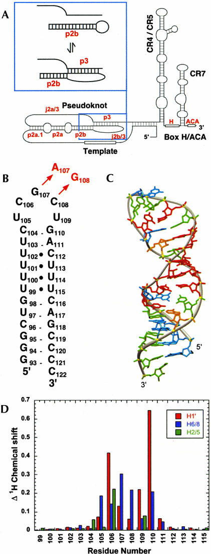
FIGURE 1.
(A) Secondary structure of the human telomerase RNA, with the domains identified as described in Chen et al. (2000). Inset: Conformational equilibrium between the P2b-P3 pseudoknot and P2b hairpin conformations. (B) Secondary structure representation of the HPwt RNA representing nt 94–120 from the wild-type P2b hairpin. The DKC mutation in the P2b helix (HPdc) is indicated in red. (C) Solution structure of the HPwt hairpin (PDB accession code: 1NA2; Theimer et al. 2003). Nucleotides are colored red (U), green (C), orange (A), and cyan (G). (D) Observed changes in chemical shift (Δppm) between HPwt and HPdc are plotted by residue number and include H1′ (red), H6/8 (blue), and H5/2 (green) resonances.

