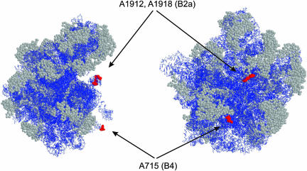
FIGURE 4.
Modeling of interfering positions into the crystal structure of Deinococcus radiodurans 50S subunits (Harms et al. 2001; PDB accession code 1KC9). The left view is from the L1 side and the right view is from the 30S side of the 50S subunit. Arrows indicate interfering positions (E. coli numbering) and corresponding bridges. RasWin molecular graphics was used to highlight interfering positions in red spacefill. The rest of the RNA is blue and α-carbons of r-proteins are in gray spacefill.

