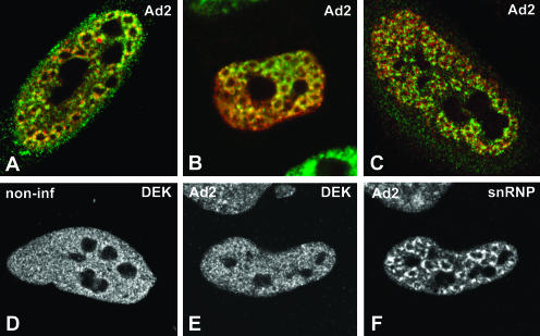
FIGURE 2.
Colocalization of EJC proteins and spliceosomal snRNPs in adenovirus-infected cells. (A–C) A superimposition of red and green images corresponding to double-labeling experiments performed in HeLa cells infected with adenovirus for 14–16 h. The distribution of EJC proteins is compared with that of spliceosomal snRNPs (A,B) and spliced viral major late mRNAs (C). (A) Cells were double labeled with antibodies directed against REF/Aly (green staining) and Sm proteins (red staining). (B) Cells expressing zzY14 (red staining) were immunolabeled with an antibody specific for the U2 snRNP B″ protein (green staining). (C) Cells were immunolabeled with anti-SRm160 antibodies (green staining) and hybridized with SJ1 probe (red staining). The distribution of DEK was analyzed by immunolabeling in noninfected (D) and infected (E) HeLa cells. The cell depicted in E was double labeled with antibody Y12, specific for Sm proteins (F).

