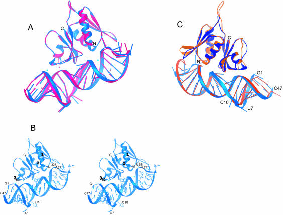
FIGURE 3.
The structure of E. coli S8/mRNA1a and its comparison with the T. thermophilus S8/helix 21 structure. (A) By using the phosphorus atoms of the S8-binding site (A12–G17/C34–U38) and the Cαs of S8, Complex A (blue) was superimposed on complex B (pink). (B) Stereoview of the structure of the E. coli S8–mRNA complex shown in blue (complex A), with the Zn2+ ions shown in cyan. (C) A comparison of E. coli complex A (blue) with T. thermophilus S8/helix 21 (orange; Wimberly et al 2000). Superposition was done by using the phosphorus atoms of the S8-binding site (A12–G17/C34–U38) and the Cα atoms of S8. Note that the complexes have been rotated 180° relative to those shown in A and B.

