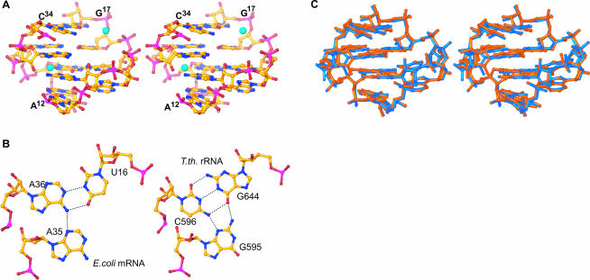
FIGURE 4.
Structure of S8s mRNA and rRNA-binding sites. (A) Stereoview of E. coli mRNA-binding site (A12–G17/C34–U38). Zn ions, shown in cyan, are tetrahedrally coordinated and participate in water-mediated interactions with the mRNA. (B) The U16–A35–A36 base triple from E. coli mRNA1a (left) and the corresponding base triple from T. thermophilus G644–C596–G595 (right). (C) Stereoview of the superposition of the E. coli mRNA-binding site (blue) with the T. thermophilus rRNA-binding site (orange). The C1′ atoms of the conserved residues, which interact with S8 (C15, G37, A14, A12), were used for the superposition.

