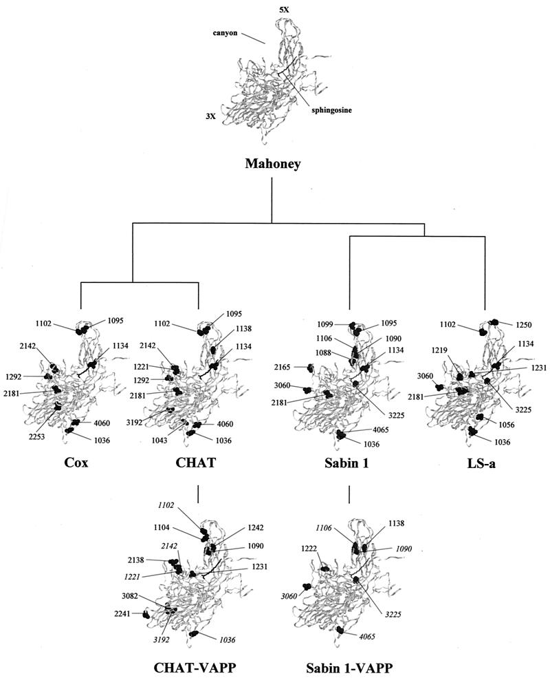
FIG. 2.
Ribbon diagrams of the α-carbon trace of the wild-type 1 poliovirus Mahoney strain protomer (21), viewed from the side of the virion (the outside is toward the top left of the image and the inside is toward the bottom right). The virus particle consists of 60 protomers, each containing a single copy of VP1, VP2, VP3, and VP4, arranged in icosahedral symmetry. 3X and 5X, threefold and fivefold axes of symmetry. The location of the canyon and the sphingosine in the hydrocarbon-binding pocket are also indicated. Amino acid mutations found in CHAT and Cox strains are shown and compared to those reported for Sabin 1 and LS-a strains (30, 48). Mutations identified in CHAT-VAPP isolates and those previously published for Sabin 1-VAPP strain 1-IIs (16) are also displayed. Reversions to wild Mahoney amino acid sequences are shown in italics.

