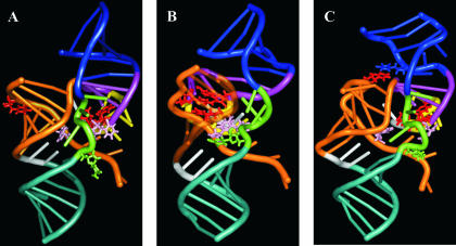
FIGURE 7.
Three-dimensional representations of the ribozyme–substrate complexes. (A) Initial ribozyme–substrate complex (RzS). (B) Active ribozyme–substrate complex (RzS′) where the U+4 of the substrate is stacked between both the C22 and U23 of the ribozyme. (C) Inactive ribozyme–substrate complex (RzS*) where the U+4 of the substrate is stacked to the U17 of the ribozyme. The P1 stem is orange, the P2 blue, the P3 purple, and the P4 cyan. The J1/4 junction is white, the J4/2 green, and the L3 loop yellow. The nucleotides U+4 of the substrate and C22 and U23 of the ribozymes are red. The U17 of the ribozyme is blue (C). The C−1 of the substrate and the U77 of the ribozyme are pink. Finally, the C76 of the ribozyme is green. Supplementary representations including a 360° rotation are available at http://132.210.163.235/jo/Figure8.html.

