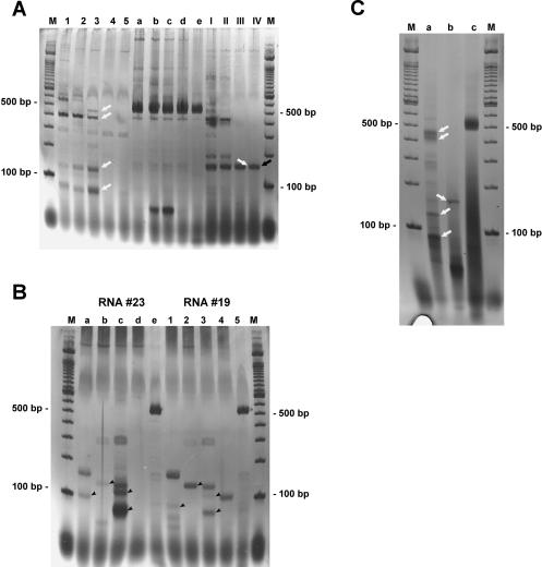
FIGURE 4.
cTnI PCR bands on a silver-stained polyacrylamide gel. (A) A hybrid PCR performed with the reverse primer pairs 363R/731R (lanes 1–5) and 419R/599R (lanes I–IV) and cDNA synthesized at different temperatures (45°C–60°C). As a control for cDNA quality, PCR with forward primer 255F and reverse primer 731R was performed in parallel (lanes a–e). Clearly visible, the complex band pattern obtained with cDNA synthesized at 45°C (lanes 1,a,I) is gradually reduced when cDNA is produced at higher temperatures (50°C, lanes 2,b,II; 55°C, lanes 3,c,III; 60°C, lanes 4,5,d,e,IV; lanes 5,e show an independent experiment at 60°C) leaving only a few bands, whereas the control PCR shows the expected band at around 500 bp with more or less the same intensity in all lanes. The product from lane IV (black arrow) was shotgun cloned and sequenced. The clone obtained showed the expected antisense/sense hybrid structure (see text). (B) A reverse PCR band pattern achieved with cTnI reverse primer pairs 419R/599R (lanes a,1), 407R/599R (lanes b,2), 363R/731R (lanes c,3), and 363R/599R (lanes d,4). cDNA was derived from different RNA preparations (called #23, lanes a–e, and #19, lanes 1–5) and synthesized at 60°C with Thermoscript (Invitrogen). Clearly, the different cDNAs give comparable band patterns with identical primer pairs, although some differences in intensity exist (arrowheads). Only PCR with 363R/599R (lanes d,4) gave no visible band in RNA #23 but one band in RNA #19. The control amplification of cTnI with the forward primer 255F and reverse primer 731R (lanes e,5) gave the expected 500 bp fragment in both cases. (C) The results of a one-tube RT-PCR with Tth-Polymerase on total RNA are shown. The RT step was done for 20 min at 60°C. (Lane a) The two reverse primer 363R and 731R had been used. (Lane b) 419R and 599R. The control PCR with the forward primer 255F and the reverse primer 731R is shown in lane c. When compared to part A of this figure, a nearly identical fragment pattern is visible (white arrows). Cloning and sequencing of this RT-PCR identified hybrid RNA as expected.

