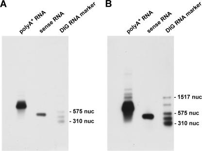
FIGURE 5.
Example of a Northern blot. Shown is the Northern blot of 1.5 μg rat poly(A)+ RNA after the incubation with cTnI hybrid RNA (clone 3.1A, DIG-marked) as probe. A and B are the same blots after different exposition times (1 min in A and 10 min in B). A prominent band at about 800 nucleotides (nuc) is visible in both cases. Also, the positive control (in vitro transcribed cTnI sense RNA) gives the expected signal at 500 nuc. After 10 min of exposition (B), several additional bands are visible that could represent hybrid RNA. As molecular weight marker the DIG-RNA marker III (Roche) was used.

