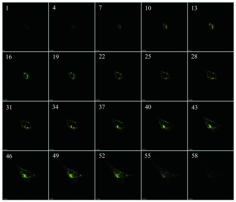
Figure 2.
Representative optical sections displaying internalization and perinuclear accumulation of antisense ODN by PFV delivery. HEK 293 cells were incubated with PFV–ODN for 4 h and analyzed by confocal laser scanning. The optical sections (total 59) were scanned incrementally along the z-axis of the cell at a space distance of 0.15 µm. Section numbers on the z-axis are given in the upper left corner.

