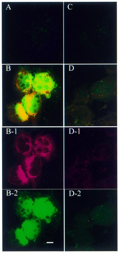
Figure 4.
Confocal microscopy projection image of cellular distribution of antisense ODN in 518A2 (A and B) and HEK 293 cells (C and D) after 48 h exposure to free antisense (A and C) or PFV-encapsulated antisense (B and D). Rh-PE label is displayed as a red fluorescent image (B-1 and D-1) and antisense ODN is displayed as a green fluorescent image (B-2 and D-2). Scale bar 10 µm.

