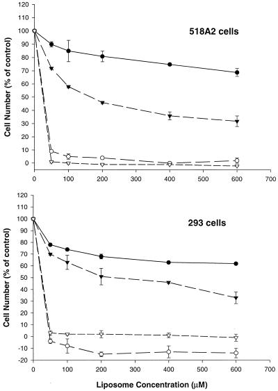
Figure 5.
Effect of time and lipid concentration on cellular viability. 518A2 and HEK 293 cells were incubated with PFV–ODN (closed circles and closed triangles) or cationic liposome–ODN complexes (open circles and open triangles) at various lipid concentrations. Cells were assessed for cytotoxicity using the XTT assay after 24 (closed and open circles) or 48 h incubation (closed and open triangles) as described in Materials and Methods. Cell viability was expressed as a percentage of surviving cells as compared to untreated control cells. Values (means ± SD) were obtained from two or three experiments and each experiment was done in triplicate.

