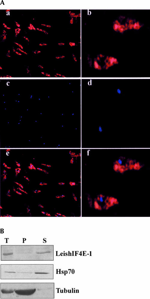
FIGURE 6.
Subcellular localization of LeishIF4E-1. (A) Localization by immunostaining. Leishmania parasites were attached to coverslips coated with poly-lysine, fixed with methanol, and reacted with rabbit antiserum raised against LeishIF4E-1 (1:4000). The fixed cells were then incubated with goat anti-rabbit antibodies conjugated with Cy3. After washing away the second antibody, the cells were stained with DAPI and mounted over glass slides. The slides were visualized in a fluorescence microscope for Cy3 (a,b) and DAPI (c,d) staining. Pictures of similar fields were overlapped to highlight the position of nuclei (e,f). (B) Localization by Western analysis of fractionated cells. Extracts were prepared from whole cells (T) and from subcellular fractions containing the cytoplasm (S) and nucleo-cytoskeletal fractions (P) and separated over 15% SDS-PAGE. Western blots were reacted with antibodies raised against recombinant LeishIF4E-1. Control blots containing equal loads of the same fractions were reacted with a monoclonal antibody against Hsp70 and α-tubulin, representing the cytoplasmic and the nonsoluble nucleo-cytoskeletal fractions, respectively.

