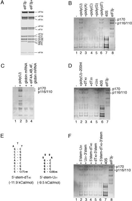
FIGURE 1.
Subunit composition of HeLa eIF3j+ and eIF3j− analyzed by electrophoresis on 4%–12% Bis-Tris NuPAGE gel using MOPS buffer system (Invitrogen), followed by Coomassie staining (A). Presence of eIF3 in the peak fraction corresponding to 40S ribosomal subunits after sucrose density gradient centrifugation, analyzed by electrophoresis on (B,D,F) 4%–12% Bis-Tris NuPAGE gel using MES buffer system (Invitrogen) or (C) SDS-11% polyacrylamide, followed by Coomassie staining. (B,F) 40S subunits (lane 7) and eIF3j− (lane 8) and (D) 40S subunits (lane 6) and eIF3j− (lane 7) were incubated in the absence (lane 1) or in the presence of poly- or oligonucleotides, as indicated, and separated by centrifugation in 10%–30% linear sucrose density gradients. (C) 40S subunits were incubated with eIF3j− alone (lane 1), eIF3j−and poly(U) (lane 2), eIF3j− and globin mRNA (lane 3) or eIF3j−, globin mRNA, eIFs 4A, 4B, and 4F (lane 4) and separated by sucrose density gradient centrifugation. eIF3 subunits are labeled to the right of panels A–D and F. (E) Sequences and structures of 5′ stem-U31 and 5′stem-dT40 oligonucleotides.

