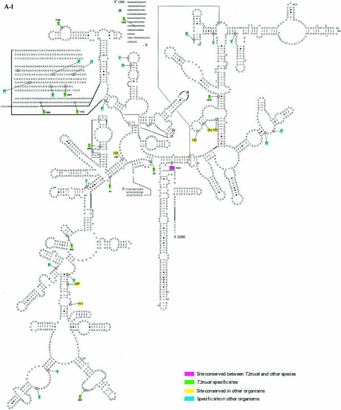
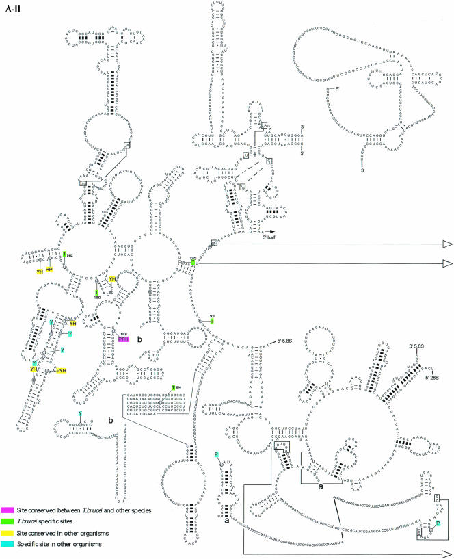
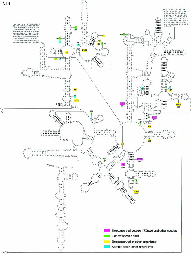
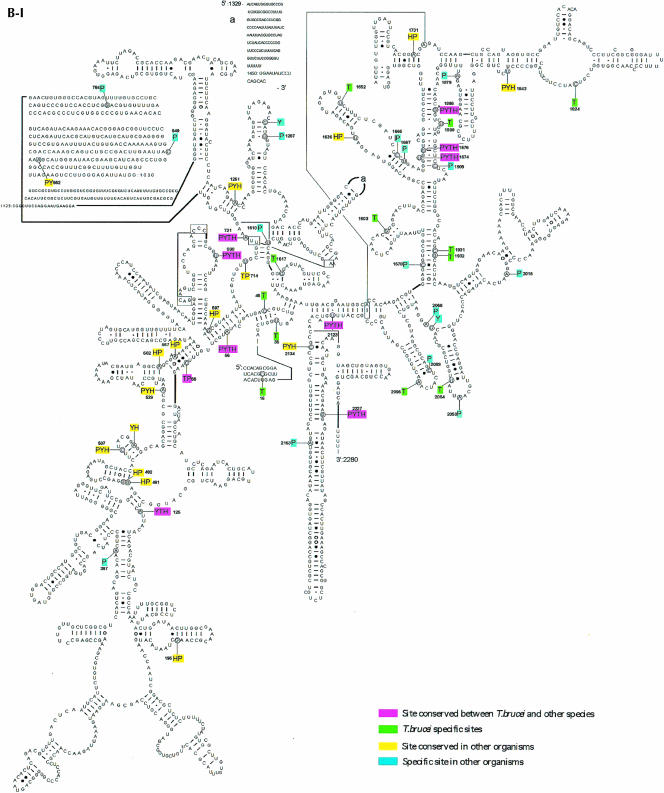
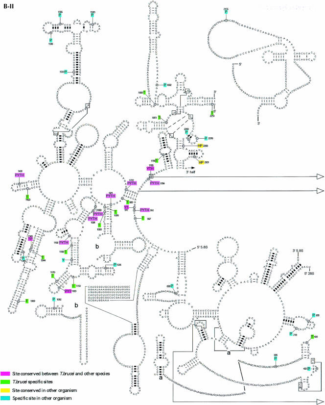
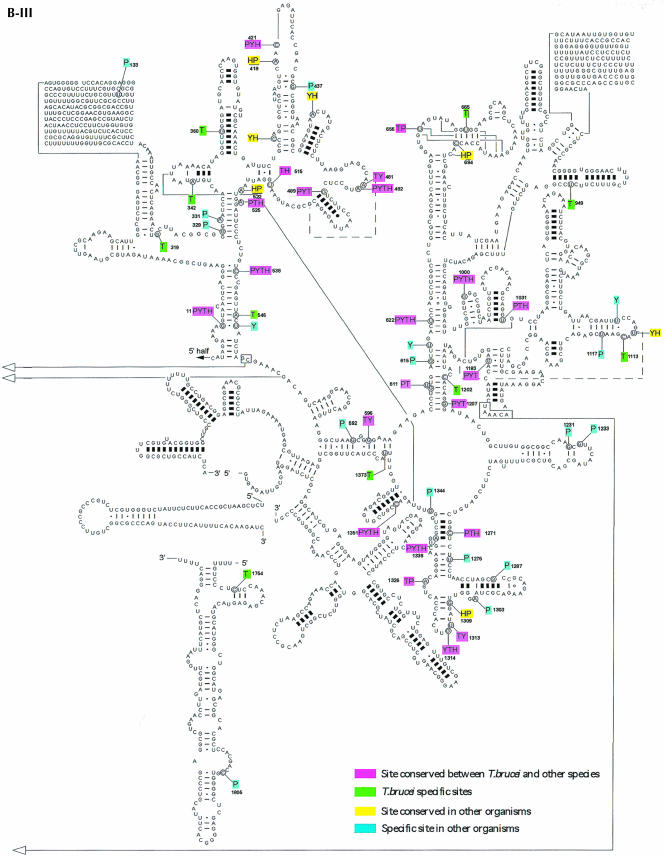
FIGURE 5.
Localization of modified nucleotides on the secondary structure of rRNA. (A) Pseudouridine on rRNA SSU (A-I), LSU 5′ half (A-II), and 3′ half (A-III). The secondary structure of T. brucei rRNA was predicted based on the structure presented at http://www.icmb.utexas.edu. The modified nucleotides in T. brucei (T), Saccharomyces cerevisiae (Y), and plant Arabidopsis (P) are indicated. The sites of humans (H) represent only the most conserved modifications. The designation of colors is indicated in each panel. (B) 2′-O-Methylations on rRNA SSU (B-I), LSU 5′ half (B-II), and 3′ half (B-III). The modified nucleotides in T. brucei (T), S. cerevisiae (Y), and plant Arabidopsis (P) are given. The highly conserved modifications in humans (H) are also shown.

