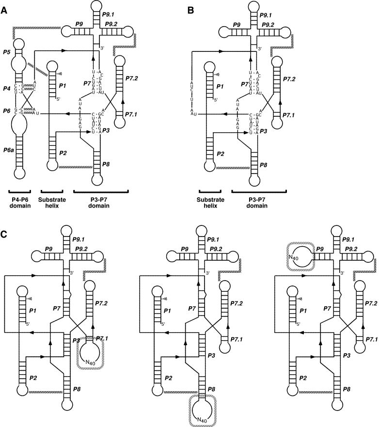
Figure 1.
Secondary structure of the ribozymes. (A) Wild-type T4 td ribozyme. (B) M1 mutant ribozyme. (C) Three sub-libraries used for the selection: (left) sub-library 7.1; (middle) sub-library 8; (right) sub-library 9. The structure model is according to Cech et al. (35). Gray arrowheads point to 5′ splice sites. Tertiary interactions are indicated by gray lines.

