
Figure 5.
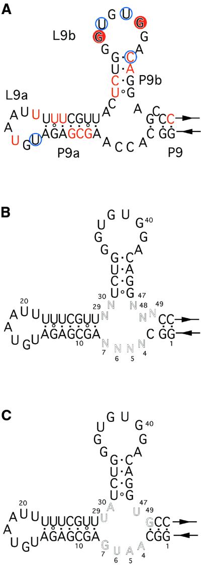
(A) Secondary structural model of the clone 2 ribozyme. The RNase V1 cleavage sites are indicated by red circles and the substrate- independent cleavage sites are shown by red letters. RNase A cleavage sites are shown by blue circles. (B) The partially randomized clone 2 library. (C) Secondary structure of the clone 2.1 ribozyme.