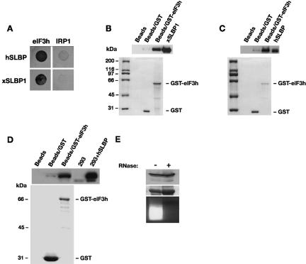
FIGURE 3.
SLBP interacts with eIF3. (A) Yeast two-hybrid assay in MaV99 strain using hSLBP or xSLBP1 fused to GAL4 DNA-binding domain (DB) and eIF3 subunit h or IRP1 fused to GAL4 activation domain (AD). IRP1 is used as a negative control. Transformed colonies were diluted and plated on selective media lacking histidine and containing 25 mM 3-AT. (B) GST-pulldown assay with GST-eIF3h fusion protein and recombinant xSLBP1. GST-eIF3h or GST bound to glutathione sepharose beads or beads alone were incubated with His-tagged xSLBP1. Samples were analyzed by Western blotting using anti-His antibody (top panel) or visualized by staining with Coomassie brilliant blue (bottom panel). xSLBP1 indicates recombinant protein input control (1/30th reaction volume). Note that GST is in excess over GST-eIF3h, indicating that the enrichment of xSLBP1 observed is due to eIF3h. (C) GST-pulldown assay with GST-eIF3h and recombinant hSLBP. The assay was performed as in B except that recombinant hSLBP was used and detected with an anti-hSLBP antibody. hSLBP indicates recombinant protein input control (1/30th reaction volume). (D) Assays were performed and analyzed as in C except that extracts from HEK293 cells expressing HA-tagged hSLBP were used. 293 and 293+ hSLBP are input controls (1/30th reaction volume). Proteins were visualized by Western analysis using an anti-hSLBP antibody (top panel) or by staining with Coomassie brilliant blue (bottom panel). (E) The interaction between eIF3h and hSLBP is not dependent on RNA. Pulldown assays were performed in parallel with D, except that one sample was pretreated with RNase A (+). hSLBP was visualized by Western blotting (top panel) and GST-eIF3h by staining with Coomassie brilliant blue (middle panel). The efficiency of the RNaseA treatment was confirmed by staining extracts with ethidium bromide after agarose gel electrophoresis (bottom panel).

