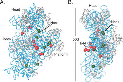
FIGURE 5.
Changes observed in the human 40S subunit due to 60S subunit association, modeled onto the crystal structure of the E. coli ribosome. (A) The small subunit shown from the 60S subunit interface side. (B) The small subunit shown from the Exit-tRNA side, where the large subunit would be to the left. The 30S ribosomal subunit from the E. coli 70S ribosome is shown with RNA in blue and proteins in gray. Positions of nucleotides that become protected from DMS modification in 80S ribosome compared with 40S subunits are indicated by red spheres; nucleotides that have enhanced reactivity in 80S monosomes are indicated by green spheres. Domains in the subunit are marked: Head, Body, Platform, h44, penultimate stem in the 3′ minor domain.

