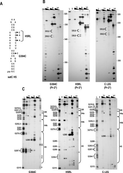
FIGURE 5.
Structural differences in the 3′ region of G304C, H5RL, and CΔ2G. (A) Location of alterations in G304C and H5RL. CΔ2G contains a deletion of the 5′ terminal two guanylates (not shown). (B) Cleavage pattern in the Pr regions of G304C, H5RL, and CΔ2G. Note the distinctive RNase A cleavages of U338/C339 and RNase T1 cleavages of G340/G341 that are similar to those of C+18b (see Fig. 3A ▶). Abbreviations above each lane are as described in the legend to Figure 3 ▶. (C) Cleavage pattern in the DR/H4a/H4b and H5 regions of G304C, H5RL, and CΔ2G. Guanylate residues in the RNase T1 ladder lane are identified by their positions, as is the prominent cleavage at U280. The boundaries of the H5, H4b, H4a, and DR regions are shown to the right. Asterisks denote cleavages not found in wild-type satC.

