FIGURE 2.
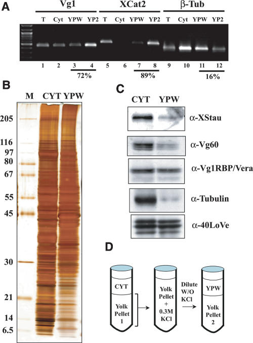
Analysis of the yolk pellet associated localization machinery. (A) RNA from a pool of 40 isolated stage III–IV oocytes was extracted from fractions prepared as illustrated in panel D and RT-PCR from 5 μg of RNA prepared from the fractions indicated was performed using primers specific to Vg1, Xcat-2, or β-Tubulin mRNA. The result demonstrates that the two vegetally localizing RNAs are enriched in the yolk pellet to a much greater extent than tubulin mRNA, and that 0.3 M KCl is sufficient to partially release them from the pellet. Quantification of the fraction of RNA present in the yolk pellet (calculated by adding YPW and YP2 and dividing by the sum of cytoplasm, YPW, and YP2) is indicated below these lanes. (B,) 30 μg of Cytosolic protein (Cyt) or Yolk Pellet Wash protein (YPW) was analyzed by 10%–15% gradient SDS-PAGE. Proteins in these lanes are from a single preparation, sequentially extracted from the same complete ovary. The conditions for extraction were identical to those in panel A. (C) An equivalent gel to that in panel B was Western blotted and probed sequentially with antibodies against several factors implicated in vegetal mRNA localization (indicated to the right of each blot) to assay their release upon washing the yolk pellet. (D) A diagram of the fractionation scheme.

