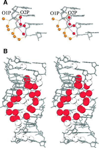
Figure 2.
Stereos showing hydration patterns in G3C3. (A) Water molecules binding to phosphates. Those H-bonded to the outer O1P are yellow, while those bonded to the inner O2P are red. (B) Major groove hydration, with the double spine of water molecules in red, and other ordered water molecules in white.

